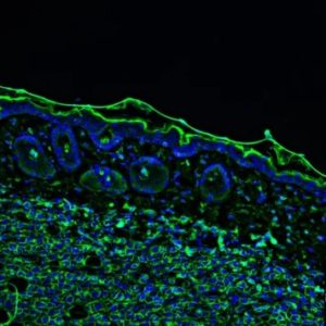
One of the most common questions we receive about CoolLED fluorescence illumination relates to the power output, which is a relic from the days where LED illumination struggled to produce the output for fluorescence microscopy. However, modern LED illumination systems now deliver more than enough light for the vast majority of applications, which in itself can be problematic unless optimised.
One of the most important questions to ask any manufacturer of custom microscopy illumination systems should instead refer to the control features available for modulating the power output, and hence the irradiance at the sample plane. The main reason for this is to protect samples against phototoxicity.
What is phototoxicity and why is it a problem?
Phototoxicity occurs mainly as a result of reactive oxygen species (ROS) released as cells are irradiated, causing damage to DNA and proteins. The most worrying aspect here is not that cells may be killed during live cell imaging, but how phototoxicity can subtly alter a cell’s physiology and skew experimental results.
Another aspect of phototoxicity is photobleaching, where the fluorophore is transiently altered into a dark, non-fluorescing state; or atoms existing in the dangerous triplet state can absorb enough energy to break covalent bonds and irreversibly damage the chemical structure, whilst also releasing ROS. The speed of photobleaching varies between fluorophores, but it can cause three problems:
-
Data misinterpretation: is signal loss due to the biology or photobleaching?
-
Time lapse experiments cut short: the observation of longer processes is limited as signal fades.
-
Inconsistent data: variable bleaching rates can impact data interpretation, for example in slide scanning and time lapse applications.
How illumination systems can minimise phototoxicity
Exactly how phototoxicity can impact cell physiology and how this can be reduced is becoming better understood all the time.1 While factors such as sample type and fluorophores come into play, the illumination technology is one of the biggest factors in optimising illumination and protecting experiments, and when creating a microscope setup these are the factors to consider:
- Less is more: The easiest step is to minimise irradiance to a level only required for image analysis, and the ability to finely control LED illumination from 0-100% makes optimisation far simpler than using ND filters with traditional lamps.
- Wavelengths: Traditional lamps tend to have a higher irradiance in the higher-energy UV region of the spectrum, which is far more damaging compared to the longer wavelength red regions. LED illumination systems offer far more choice in excitation wavelengths, with our technology currently extending to 850 nm. New fluorophores excited by these longer wavelengths are becoming available all the time, alongside the optical filters to match. Moreover, individual channel switching means the sample is exposed only to the required wavelengths.
- High-speed electronic control: Traditional lamps use mechanical shutters, and even when triggered electronically still create a lag (and potentially vibration) which can be hundreds of milliseconds. A descriptive term for this is ‘illumination overhead’ and is defined by the authors as: “the time fluorescent samples are exposed to incident light, but fluorescence emission is not being collected by the detector”.2 To drastically reduce illumination overhead, LED illumination systems with a global TTL-in can be synchronised directly to cameras featuring a TTL-out, and this can be as fast as <7 µs. This approach is especially valuable over the course of time lapse experiments and can result in improved cell health and extended studies due to reduced photobleaching. These speeds have the added benefit of increasing temporal resolution and accuracy of ratiometric measurements, and for further information please see our in-depth review of hardware options which compares speed, contrast and budget requirements.
- Pulsed illumination: The theory here is that constant illumination continues to create atoms in the fluorophore which exist in the dangerous triplet excited state. When light is instead delivered as a pulse with microsecond gaps, these triplet states have time to reset instead of damaging the fluorophore structure. Incredibly, this has been shown to achieve a 9-fold reduction of EGFP photobleaching.3
Fast control of LED illumination means the sample is only exposed to the optimum level of light at the required wavelength, and only when required.
Summary
Unfortunately, there is no ‘one size fits all’ approach and it is up to the end user to consider cell health as a vital aspect of every experiment, building in the necessary controls. However, CoolLED custom microscopy illumination systems can provide the first step, equipping them with the ability to optimise the illumination and protect both fixed and live samples.
Find out how our LED illumination technology can give you the competitive edge with data quality
Discover what it’s like working with CoolLED
If you have any questions, please get in touch
References
-
Icha, J., Weber, M., Waters, J. C, & Norden, C. (2017). Phototoxicity in live fluorescence microscopy, and how to avoid it. BioEssays, 39, . doi: 10.1002/bies.201700003
-
Tinevez JY, Dragavon J, Baba-Aissa L, Roux P, et al. 2012. A quantitative method for measuring phototoxicity of a live cell imaging microscope. Methods Enzymol 506: 291–309.
-
Kiepas, A., et al. (2020). Optimizing live-cell fluorescence imaging conditions to minimize phototoxicity. Journal of cell science, 133(4), jcs242834. https://doi.org/10.1242/jcs.242834


