For live cell imaging, label-free microscopy is a popular method which comes with several advantages. Unlike fluorescence microscopy, there are no labels that could potentially interfere with the phenomenon you want to observe. It also requires lower light levels, which is great for reducing photodamage. Happy cells will behave closer to real life, and this in turn improves confidence in experimental data. It’s also ideal for studying longer processes, with time lapse studies being extended as cells remain healthy for longer, and there is no interference from troublesome photobleaching.
On the other hand, this method is limited to observing membranes rather than molecules. And this is where fluorescence microscopy steps in. For the best of both worlds, the Nanolive 3D Cell Explorer-fluo microscope enables both label-free imaging and fluorescence microscopy, combining high quality holotomographic data with molecular data from fluorescent markers. This allows scientists to acquire two distinct types of information and perform a broad range of live cell imaging experiments on a single microscope.

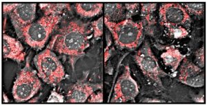
11 hr time-lapse of mouse pre-adipocytes. Mitochondria labelled with mitoTracker. Holotomographic image taken every 15 seconds; fluorescence image every 5 minutes. (Image: Nanolive)
Benefiting from custom microscopy illumination
For fluorescence microscopy illumination, Nanolive has relied on a specialised pE-300ultra Amora variant for several years. After meeting at ASCB in winter 2016, we supplied a prototype, which was integrated and tested over February 2017. The first Nanolive microscope with a CoolLED Custom Microscopy Illumination System was sold just later that year – and the rest is history!
The CoolLED Custom Microscopy Illumination System is a broad spectrum three-channel system, and in addition to the control options available, Nanolive also enjoys reliability, uniform illumination – and expert support from our CoolLED team.
You can find out more in the interview below, Head of Quantitative Biology at Nanolive, Dr Mathieu Frechin, explains the technology in more detail and gives an insight into where label-free technology is headed next.


